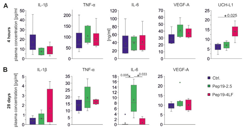
Figure 5.
Plasma levels of IL-1β, TNF-α, IL-6, VEGF-A and UCH-L1 four hours (A) and 28 days (B) after CA-CPR. Measurements are shown in pg/mL (except IL-6 after 4 h in ng/mL). Plasma levels were assessed in each group and presented as boxplots showing the quartiles, the 5th and 95th percentiles (whiskers), the median (line) and the mean (x). (A) Four hours after CA-CPR, plasma levels of cytokines and VEGF-A were measured at an elevated level compared with 28 days after CA-CPR (B). Differences in the comparison of the groups (each n = 6, unless otherwise specified) could not be detected with regard to cytokines (IL-1β: p = 0.109; TNF-α: p = 0.423; IL-6: p = 0.519) and VEGF-A (p = 0.076). UCH-L1 levels in the Pep19-4LF treated group (n = 5) were significantly higher than in the control group (n = 4, p = 0.025). (B) All plasma levels of cytokines and VEGF-A 28 days after CA-CPR were normally low. For IL-6, the group of mice treated with peptide Pep19-2.5 showed significantly higher concentration after CA-CPR (p = 0.003; pairwise: Ctrl. (n = 8) vs. Pep19-2.5 (n = 7), p = 0.005; Pep19-4LF (n = 4) vs. Pep19-2.5: p = 0.033; Ctrl. vs. Pep19-4LF: p = 0.864).

