Figure 3.
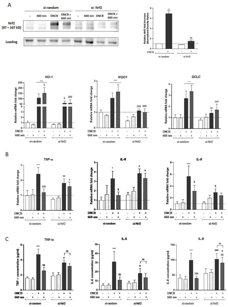
The anti-inflammatory effect of red light in KCs is Nrf2-dependent. KCs were transfected for 48 h by si-Nrf2 or si-random, and then treated with DNCB and exposed to red light. (A) Nrf2 accumulation in the knockdown model was assessed by Western blot 3 h after treatment. The blot is representative of 3 independent experiments. Histograms correspond to densitometric analysis relative to untreated si-random and are adjusted to the stain-free blot. Quantifying the expression of Nrf2 target genes encoding HO-1, NQO1, and GCLC in transfected KCs were assessed using RT-qPCR 3 h after treatment. Induction values were the ratio between gene expressions in treated cells versus gene expression in untreated si-random control. (B) Pro-inflammatory cytokines gene expression: TNF-α, IL-6 and IL-8, were assessed by RT-qPCR, 3 h after light treatment. Results are expressed as fold change compared to the untreated si-random control. (C) The inflammatory cytokines level was determined by Meso Scaled Discovery technology in the supernatant of KCs after 6 h of exposure to DNCB with or without red light illumination. Reported data are mean ± SEM of 6 independent experiments. Two-Way ANOVA followed by Tukey post hoc test, * p < 0.05; ** p < 0.01; *** p < 0.001 vs. untreated si-random; $ p < 0.05; $$ p < 0.01; $$$ p < 0.001 vs. DNCB-treated si-random, $$′ p < 0.01; $$$′ p < 0.001 vs. DNCB + 660 nm-treated si-random; # p < 0.05, ## p < 0.01, ### p < 0.001 vs. untreated si-Nrf2. ns means non-significant different.

