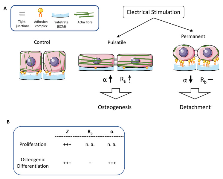
Figure 7.
Schematic presentation of morphological alterations after pulsatile and continuous electrical stimulation. (A) Pulsatile stimulation resulted in decreased space between the cellular membrane and substrate and increased α values. It appears that the cells nestle against the ground, causing the actin cytoskeleton to stiffen and the number of basal adhesion proteins to decrease. In contrast, the continuous stimulation causes the cells to round up, accompanied by increased expression of the adhesion molecules in the periphery to prevent the cells from detaching. The actin fibers mainly localize to the cell periphery. (B) Summary of the alterations of impedance (Z), resistance (Rb), and cleft impedance (α) in comparison of proliferating and osteogenic differentiating stem cells. + low increase, +++ significant increase, n. a. no alterations.

