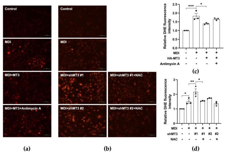
Figure 7.
MT3 impedes ROS production in the early stages of 3T3−L1 adipocyte differentiation. (a,b) 3T3−L1 cells were transfected with HA−MT3 plasmid or shMT3 plasmid (0.2 μg) prior to contact inhibition and cultured in differentiation medium with 25 nM antimycin A or 7.5 mM NAC for 12 h. ROS levels are measured by DHE assay. Representative images of 3T3-L1 adipocytes were captured at 200× magnification. Scale bar = 100 μm. MDI, a mixture of 3-isobutyl-1-methylxanthine, dexamethasone, and insulin in differentiation medium. (c,d) The quantitative analysis of the relative DHE fluorescence intensity indicated the differences in each group. The data are presented as the means ± SEM. * p < 0.05, ** p < 0.01, and *** p < 0.001 were calculated by one-way ANOVA followed by Tukey’s Test.

