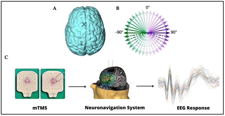
Figure 1.
(A) The stimulation site on the left pre-SMA is indicated on the brain with a cross. (B) The 36 stimulation orientations are represented as green–purple vectors, 0° corresponds to stimulation delivered in the posterior–anterior direction, whereas −180° corresponds to stimulation delivered in the anterior–posterior direction [28]. (C) Instruments and techniques used. A 2-coil mTMS transducer, tracked with a neuronavigation system, was used in combination with a 64-channel EEG recording.

