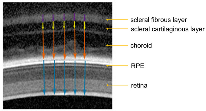
Figure 2.
Illustration of how the chicken retinal, choroidal and scleral thickness were determined in OCT images. Blue arrows: retinal thickness; orange arrows: choroidal thickness; yellow arrows: scleral cartilaginous layer thickness; purple arrows: scleral fibrous layer thickness. RPE: retinal pigment epithelium.

