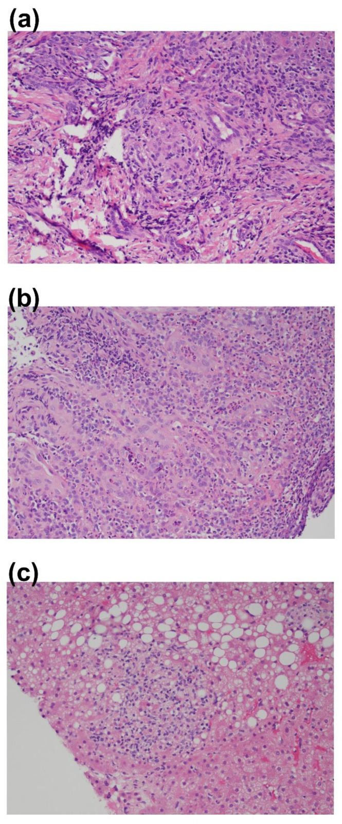
Figure 2.
Histopathological evidence of granulomatous inflammation. (a) Mucosal ulceration with focal granulomatous inflammation, hyperparakeratosis, and acanthosis: image is focused on gingival tissue at the large zone of ulceration. Within fibrous connective tissue, a heavy mixed inflammatory cellular infiltrate that is predominantly neutrophils, lymphocytes, and histiocytes is seen. Widely scattered ill-defined clusters of histiocytes and multinucleated giant cells are noted. (b) Biopsy of transverse colon tissue showing large bowel mucosa with few granulomas. No significant crypt distortion. (c) Biopsy of the liver performed via needle biopsy. Hepatic architecture shows subtle distortion with nodular areas of widened alternating with areas of narrowed liver cell plates. No significant portal inflammation or interface hepatitis. Many of the portal areas lack a distinctive portal vein, and there are occasional foci of lobular inflammation. There is mild focal steatosis, involving less than 5% of parenchyma.

