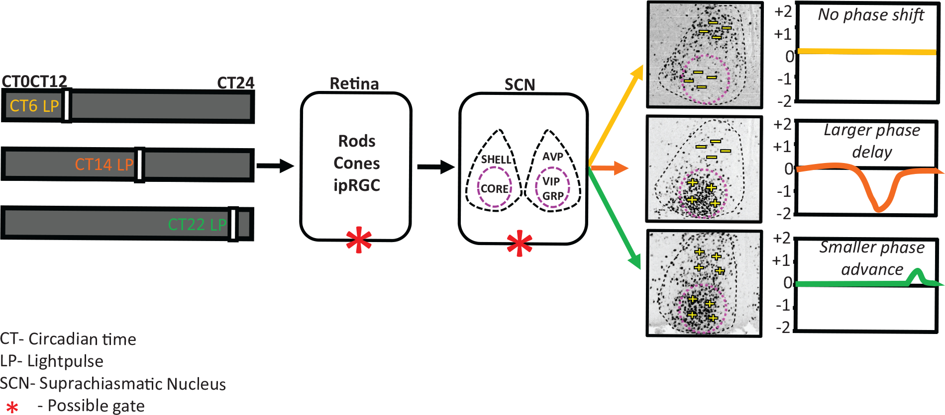
Figure 5.

Model representing light at a different time of the day differentially alters the SCN state dynamics and outputs. light input is received daily by specialized photoreceptor cells in the retina, intrinsically photosensitive retinal ganglion cells (ipRGCs), and transmitted via the retino-hypothalamic tract to the central clock located in the SCN, entraining it to the external light-dark cycle. The SCN is a bilateral structure and can be organized into 2 functionally distinct compartments: shell and core. The SCN shell contains a dense population of arginine vasopressin (AVP) neurons, and the SCN core contains vasoactive intestinal polypeptide (VIP) and gastrin-releasing peptide (GRP) neurons. Depending of the time at which the light pulse was given, the mice will show either no change or a phase shift of the activity rhythm after the pulse. There is also a clear phase-dependent response in the core and shell of the SCN at the phases when light can induce a phase shift. light pulse applied during the subjective day (CT6) causes no induction of cFos cells in the entire SCN, and no phase shift was observed. A light pulse applied at the beginning of the subjective night (CT14) causes a huge induction of cFos, but only in the core of the SCN, and larger phase delays were observed in activity. light pulses applied at the end of the subjective night (CT22) result in induction of cFos-positive cells in both the core and shell of the SCN and produce smaller phase advances. We propose that this light-signaling responsiveness is gate dependent and that the gating can be either at the retina level or within the SCN.
