Figure 1.
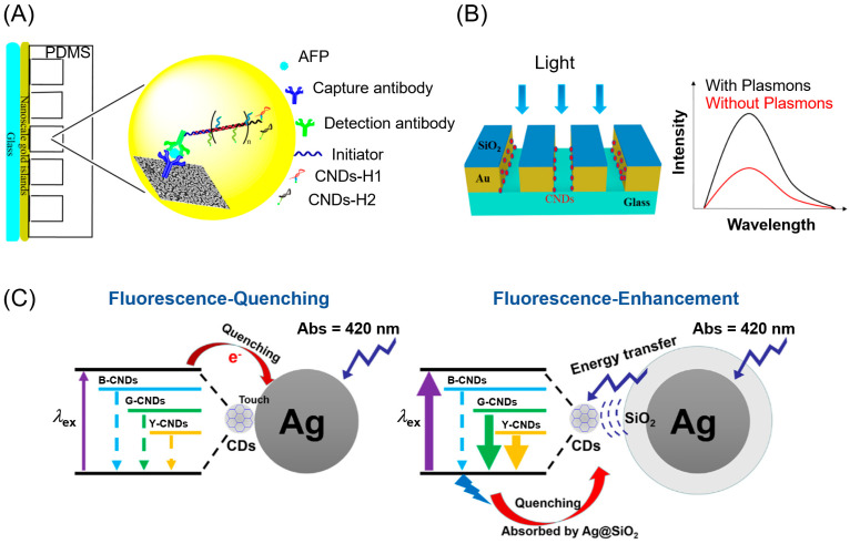
(A) Schematic illustration for the detection of AFP with Immuno-HCR and MEF of CNDs [36]. (B) Illustration of self-assembled monolayer formation and CND immobilization on a gold nanoslit surface [29]. (C) Electron transition process between CNDs and Ag or Ag@SiO2 NP for quenching (left) and enhancement (right) of fluorescence [39]. Reprinted with permission from [29,36,39].

