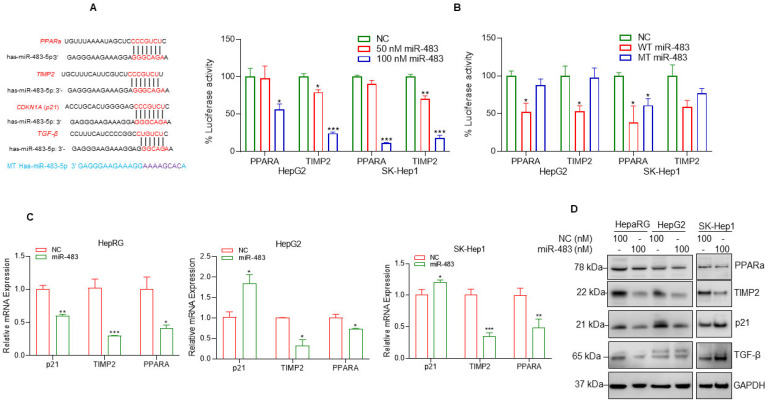
Figure 6.
miR-483 targets PPARa and TIMP2 UTRs and inhibits their expression. (A) The binding sites of miR-483 in the 3′UTR of PPARA (5), TIMP2, and p21 gene were analyzed by TargetScan (http://www.targetscan.org/vert_72/, accessed on 12 August 2022) and presented. The binding site of miR-483 in the 3′UTR of TGFB was also presented. We also generated custom-made wild-type miR-483 and mutated miR-483 nucleotide sequences and presented them. (B) Luciferase reporter assay: HCC HepG2 and SK-Hep1 cells were transfected with PPARA-3′UTR-Luciferase reporter construct (0.5 µg DNA), or TIMP2-3′UTR-Luciferase reporter construct (0.5 µg DNA) for 18 h and then cells were transfected with NC mimic or wild-type-miR-483 (50 nM and 100 nM; left panel) or wild-type-miR-483 or mutant-miR-483 (100 nM; right panel) for an additional 24 h. Transfected cells were lysed, and luciferase activity was measured. Results are representative of three independent experiments. * p < 0.05, ** p < 0.01, *** p < 0.001 compared with NC transfected cells. (C) HepaRG, HepG2, and SK-Hep1 cells were transfected with NC or miR-483 mimic (100 nM) for 30 h, and the effect of NC or miR-483 mimic on p21, TIMP2, and PPARa gene expression was analyzed by RT/qPCR. * p < 0.05, ** p < 0.01, *** p < 0.001 compared with NC-transfected cells. (D) HepaRG, HepG2, and SK-Hep-1 cells were transfected with NC mimic or miR-483 mimic for 30 h, and expression of endogenous p21, TIMP2, PPARa, and TGFβ protein expression was analyzed by western blotting. The uncropped blots are shown in Supplementary Materials.

