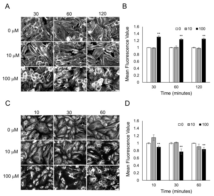
Figure 4.
Lyral exposure causes F-actin assembly and microtubule disassembly in HUVECs. Fluorescent images of F-actin phalloidin staining (A) and tubulin staining (C) after HUVECs were treated with 0, 10, 100 μM lyral for 10–120 min. Scale bar = 50 μm. Quantitative mean fluorescence intensity of F-actin staining (B) and tubulin staining (D) was measured using Fiji. A two-tailed t-test was performed to compare each lyral-treated group to the control group completed at the same time point; ** p < 0.01 as indicated.

