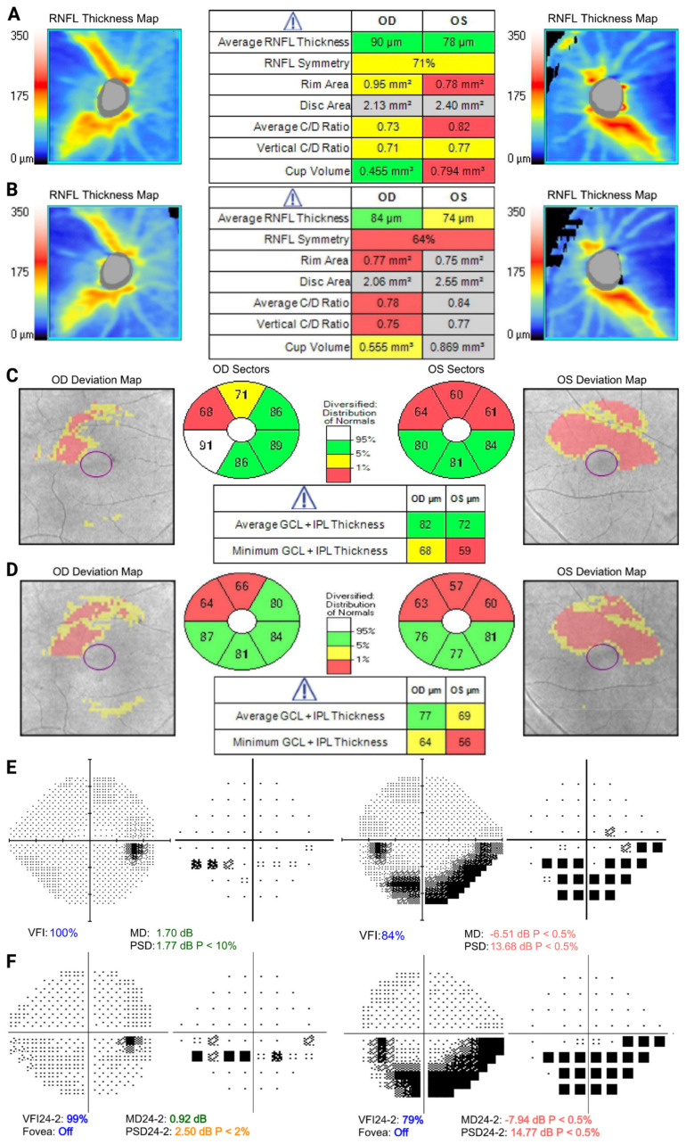
Figure 1.
Optical coherence tomography (OCT) and visual field examination of patient 1 at baseline (top) and 5-year follow-up (bottom). (A) Retinal nerve fiber layer demonstrates stable mild right inferior thinning and more pronounced left superior thinning, with progression more pronounced in the left eye over time (B). (C,D) Bilateral superior–temporal ganglion–cell complex defects, worse in the left eye. (E) Visual field shows a mild early inferior nasal arcuate defect in the right eye and a denser inferior arcuate in the left eye, which progressed over time (F).

