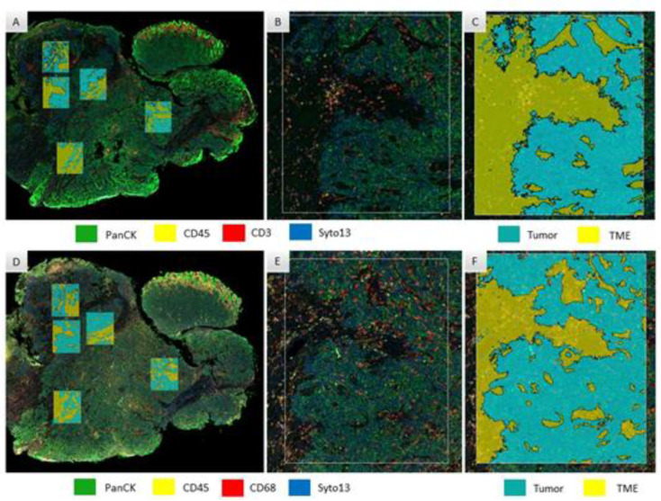
Figure 1.
Region of interest selection and segmentation for protein (A–C) and RNA (D–F) digital spatial profiler assay in an anal carcinoma sample. All the areas selected for protein assay were matched for RNA assay, here seen with low magnification in (A,D), respectively; and at high magnification for one of the regions of interest in (B,E), respectively (colors for the immunofluorescence morphology staining are seen in rectangles on the bottom of these panels). (C,F) show tissue segmentation in the same high magnification area, for tumor and tumor microenvironment compartments (colors indicating mark-up areas for tumor and TME are seen on the bottom of these panels).

