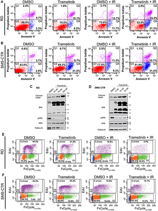
Figure 2. Combining low-dose MEK inhibition with IR results in increased apoptosis and a G1 cell cycle arrest in FN-RMS.
A. Flow cytometry plots showing propidium iodide vs. annexin V staining of DMSO- and trametinib-treated RD cells after non-IR and IR conditions. Q4 represents cells undergoing early apoptosis, whereas Q2 represents cells undergoing late apoptosis. Q3 represents live cells not undergoing apoptosis.
B. Flow cytometry plots showing propidium iodide vs. annexin V staining of DMSO- and trametinib-treated SMS-CTR cells after non-IR and IR conditions.
C, D. Cleaved PARP, BIM, pERK, and total ERK protein expression immunoblots in DMSO- and trametinib-treated RD (C) and SMS-CTR (D) cells 96hpIR. Increased trametinib doses were assessed in both cell lines.
E. Flow cytometry plots of EdU vs. DAPI staining in DMSO- and trametinib-treated RD cells in non-IR and IR conditions.
F. Flow cytometry plots of EdU vs. DAPI staining in DMSO- and trametinib-treated SMS-CTR cells in non-IR and IR conditions.

