Figure 3.
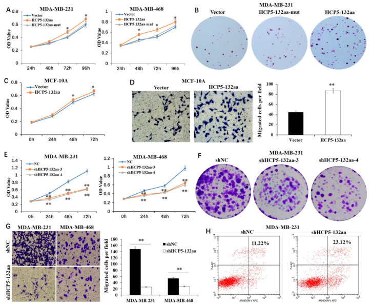
The HCP5-132aa promoted malignant phenotypes of TNBC cell lines. (A) MDA-MB-231 and MDA-MB-468 cells were transfected with the lentivirus vector, HCP5-132aa ORF or HCP5-132aa ORF-mut plasmids for the indicated times, and the cell numbers were measured by CCK8 (n = 3). (B) MDA-MB-231 cells were transfected with the indicated constructs, and their colony-forming abilities were measured after 2 weeks (×1). (C) MCF-10A cells were transfected with the vector or HCP5-132aa ORF plasmids for the indicated times, and the cell numbers were measured by CCK8 (n = 3). (D) MCF-10A cells were transfected with the vector or HCP5-132aa ORF plasmids, and migration abilities were determined using Transwell assays (×400). (E) MDA-MB-231 and MDA-MB-468 cells were transfected with the lentivirus negative control, shRNA-3, or shRNA-4 for the indicated times, and the cell numbers were measured by CCK8 (n = 3). (F) MDA-MB-231 cells were transfected with the indicated constructs, and their colony-forming abilities were measured after 2 weeks (×1). (G) MDA-MB-231 and MDA-MB-468 cells were transfected with the lentivirus negative control or shHCP5-132aa (shRNA-4), and migration abilities were determined using Transwell assays (×400). (H) MDA-MB-231 cells were transfected with the lentivirus negative control or shHCP5-132aa, and the percentage of apoptosis cells were measured by flow cytometry. * p < 0.05, ** p < 0.01.

