Figure 5.
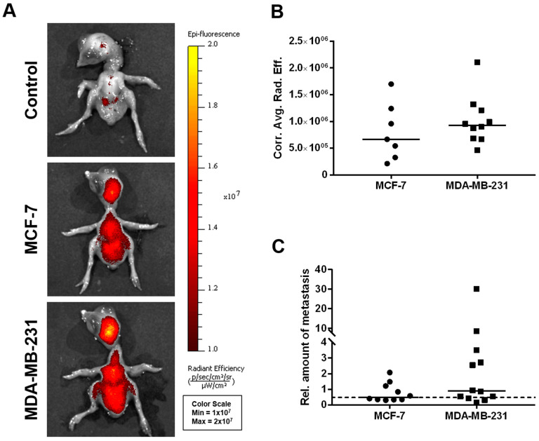
In vivo dissemination capability of MCF-7 and MDA-MB-231 breast cancer cells in the CAM xenograft assay. (A) Representative images of chicken embryos as obtained by fluorescence imaging 5 days post-engraftment of deep-red fluorescence-labeled MCF-7 and MDA-MB-231 breast cancer cell pellets onto the CAM (unlabeled cells of both the MCF-7 and MDA-MB-231 cell lines served as controls). (B) Corrected average radiant efficiency as assessed by fluorescence imaging in chicken embryos to determine the in vivo dissemination potential of MCF-7 (n = 7) and MDA-MB-231 (n = 10) cells. (C) Relative amount of disseminated tumor cells within the livers of chicken embryos as determined by human-specific Alu qPCR 5 days after engraftment of the breast cancer cell lines MCF-7 (n = 10) and MDA-MB-231 (n = 12). A value of 1 serves as a reference and indicates an amount of 0.01 ng/mL of human genomic DNA. The dotted line represents the cutoff for tumor cell dissemination detection, which was defined by the sensitivity limit of the Alu qPCR method. Medians of all data are presented as lines in the graphs. For statistical analysis, the Mann-Whitney test was used; the post-analysis was performed using Dunn’s multiple comparison tests.

