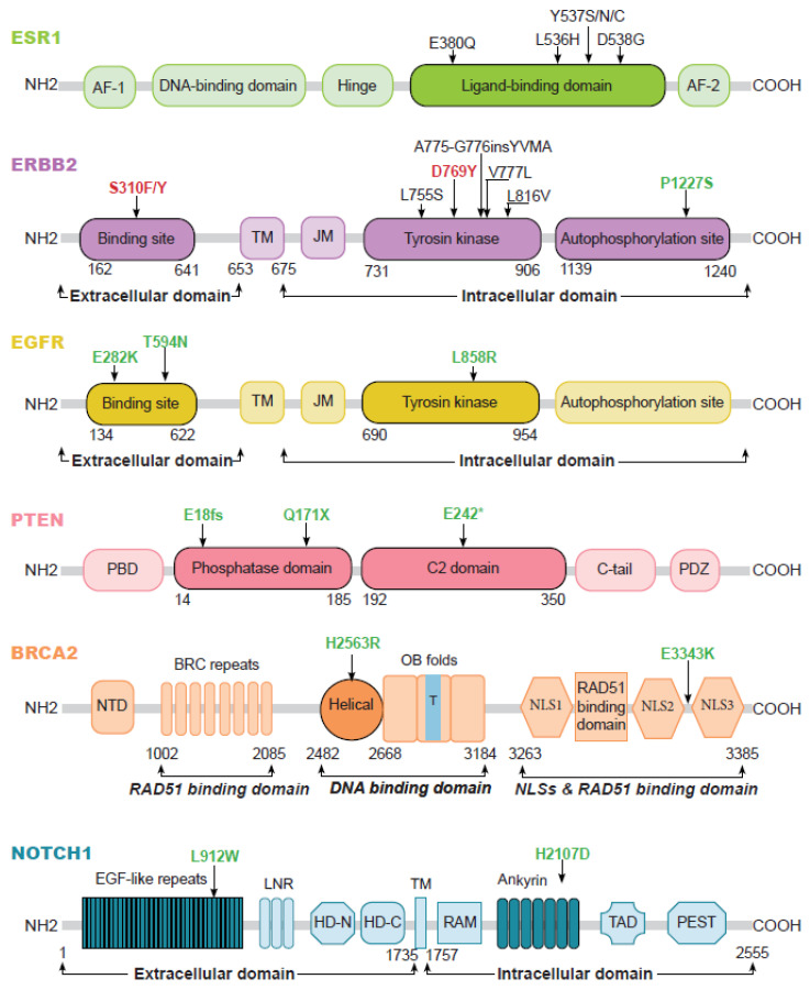
Figure 3.
Cartography of the mutations reported in the meta-analysis for the 6 genes with an increased mutation prevalence in brain metastases. Mutations exclusive to brain metastases are identified in green. Those exclusive to extracerebral metastases are in red, and the mutations common to both sites are in black. AF-1: activation function-1, AF-2: activation function-2, TM: transmembrane, JM: juxtamembrane, PBD: PIP2-binding domain; NTD: N-terminal domain, OB folds: oligonucleotide binding folds, T: tower domain, NLS: nuclear localization sequence, EGF-like repeats: epidermal growth factor-like repeats, LNR: Lin12/Notch repeat, HD-N: heterodimerization domain- N terminal, HD-C: heterodimerization domain- C terminal, RAM: Rbp-associated molecule, TAD: transactivation domain, PEST: a region rich in prolone (P), glutamate (E), serine (S) and threonine (T), *: means there is a codon stop.

