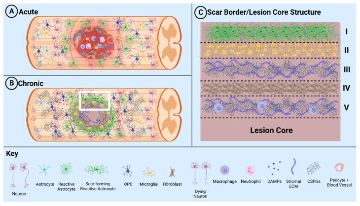
Figure 1.
Schematic diagram illustrating glial scar formation. (A) Formation of the lesion and scar during the acute phase of SCI. (B) Formation of the fibrotic tissue and glial scar during the chronic phase of SCI. (C) White box region in B detailing the layers of the glial scar: I. astrocytes, II. microglia, III. secreted stromal ECM/CSPGs, IV. fibroblasts, and V. stromal ECM/CSPGs and penetrating macrophages.

