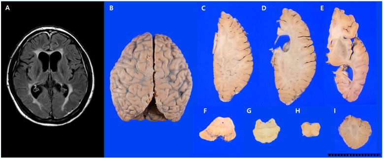
Figure 1.
Brain magnetic resonance imaging (MRI) and gross findings of the brain in case 1. (A) Brain MRI (FLAIR sequence) revealed high-signal intensities in the bilateral periventricular white matter. (B) Mild gyral atrophy of the bilateral frontal and parietal lobes. (C–E) Horizontal sections of the right hemisphere revealed white matter degeneration of the frontal and parietal lobes, sparing of subcortical U-fibers, and severe corpus callosal atrophy. (F–H) There was symmetrical atrophy of the cerebral peduncle and pyramid and (I) a relatively preserved cerebellum.

