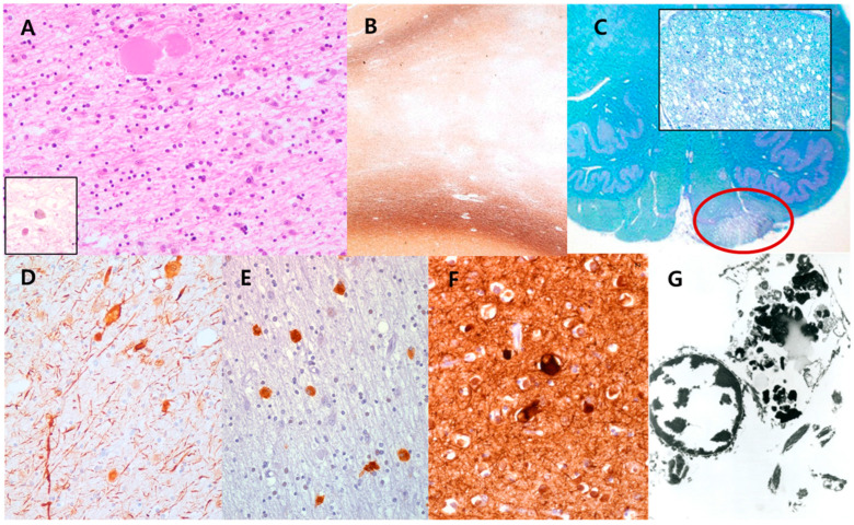
Figure 2.
Histopathological findings of the brain in case 1. (A) The white matter lesions revealed scattered axonal spheroids and pigmented macrophages (inset) on hematoxylin-eosin stain (200×), and (B) extensive loss of myelinated axons with sparing of subcortical U-fibers on Bielschovsky’s silver staining (40×). (C) The unilateral corticospinal tract (red circle) was also affected by the loss of myelinated axons or pigmented macrophages (LFB, scan view; inset: LFB, 200×). Immunohistochemically, (D) axonal spheroids and pigmented macrophages were positive for phosphorylated neurofilaments (NF) (200×) and (E) CD68 (400×), respectively. (F) A few ballooned cortical neurons were stained with phosphorylated NF (400×). (G) Ultrastructurally, the pigmented granules in macrophages exhibited lipofuscin ceroids (7000×).

