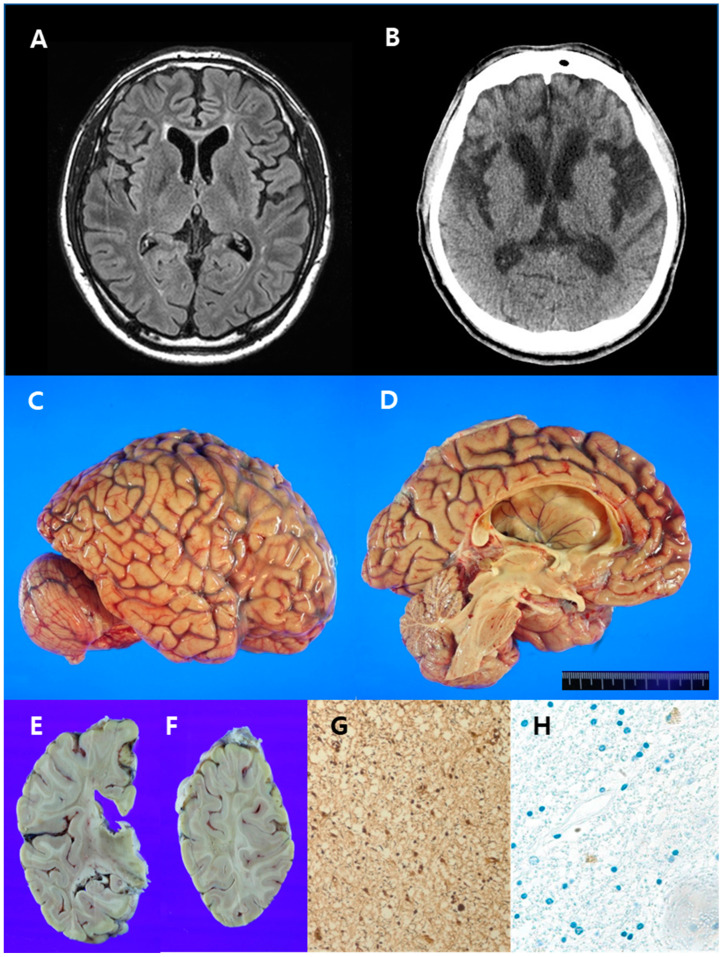
Figure 3.
Radiological images, gross and histopathological findings of the brain in case 2. (A) Brain magnetic resonance imaging (FLAIR sequence) revealed high-signal intensities in the white matter around the anterior horns of the bilateral lateral ventricle. (B) Brain computed tomography was performed six years after the initial brain MRI and revealed hypodense lesions in the white matter around the anterior horns of the bilateral lateral ventricles and mild brain atrophy with hydrocephalus ex vacuo. (C,D) Findings included moderate gyral atrophy of the frontal lobe, mild gyral atrophy of the parietal lobe, marked atrophy of the corpus callosum, and hydrocephalus ex vacuo. (E,F) The white matter was severely degenerated in the frontal, parietal, and temporal lobes and in a focal area of the occipital lobe. Subcortical U-fibers were spared. (G) Severe axonal loss, but absent axonal spheroids, was noted (Bielschovsky’s silver stain, 200×). (H) Pigmented macrophages were sparsely observed (CD68, 200×).

