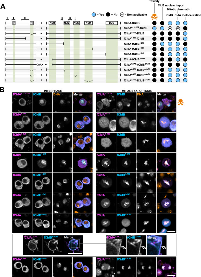
Fig 5. Phenotypes resulting from CidA-CidB mutants co-expression in cellulo.
(A) Schematic drawing of fCidA and fCidB wild-type and mutant combinations co-expressed with the T2A self-cleaving peptide under the pAc5 promoter. The resulting phenotypes presented on the right include the toxicity (i.e. no mitosis and apoptotic events observed), the CidB nuclear import during interphase, and the CidA and CidB mitotic localizations (i.e. on the chromatin). (B) Confocal images of cells expressing the constructs presented in (A) in interphase and mitosis or apoptosis -skull and crossbones symbol-. Images of CidBΔNuf1- CidACAAX co-expression are presented separately in a box with an enlargement on the right. CidA in magenta, CidB in cyan and DNA -Hoechst- in yellow. Scale bar = 10 μm.

