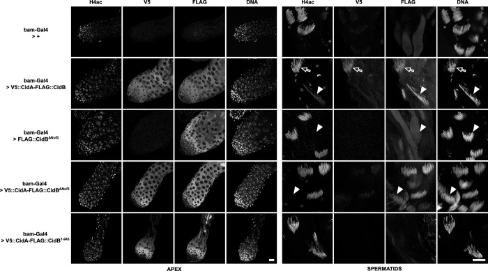
Fig 6. Spatial-temporal localizations of Cid effectors in transgenic D. melanogaster testes.
Confocal images of D. melanogaster testes of the indicated genotypes. Apical region with proliferative germ cells is shown on the left and canoe stage spermatids on the right. The immunofluorescence stainings reveal from left to right for each zone of the testis the acetylated histone 4 -H4ac- lost during the histone-to-protamine transition, V5::CidA, Flag::CidB, and DNA stained with DAPI. Arrows indicate cysts positive for H4ac before the histone-to-protamine transition while arrowheads point toward post-transition cysts negative for H4ac. The first horizontal row is a negative control testis which only expresses bam-Gal4. The testis shown in the second row expresses wild-type tCidA and tCidB. Representative spermatid cysts still positive for histones (H4ac) are indicated with an arrow. Arrowheads show cysts after the histone-to-protamine transition. Scale bars = 20 μm.

