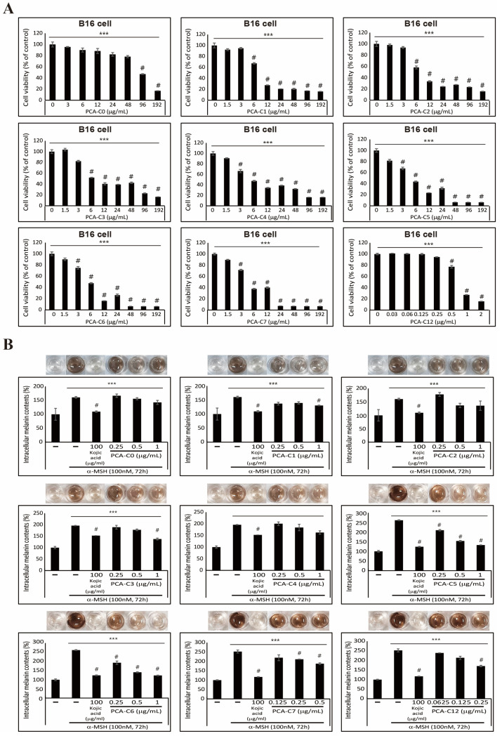
Figure 2.
PCA derivatives inhibit α-MSH-induced melanin synthesis in B16 melanoma cells. (A) Cell viability results showed the inhibition of B16 cell proliferation after treatment with an increased PCA-C(X) concentration for 24 h. The data represent three independent tests. # p < 0.001 versus control. *** p < 0.001 (ANOVA test). (B) Intracellular melanin contents were determined using cultured media containing secreted melanin after α-MSH or/and PCA-C(X) treatment for 72 h. The results of extracellular melanin content can be found in Table S1. The pictures show the color of the culture medium, and the melanin content was reduced by PCA-C(X). The data are representative of three independent experiments. # p < 0.001 versus control. *** p < 0.001 (ANOVA test).

