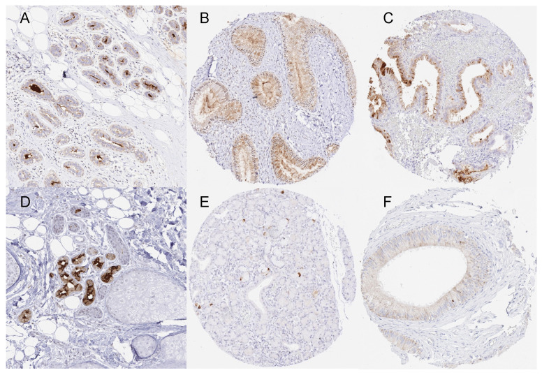
Figure 1.
Mammaglobin-A immunostaining of normal tissues. The panels show an apical membranous and cytoplasmic staining of variable intensities in luminal cells of the breast ((A), magnification from a TMA spot), endocervical glands (B), endometrial glands (C), eccrine glands of the skin ((D), magnification from a TMA spot), scattered epithelial cells of submandibular gland (E), and some chief cells in the corpus epididymis (F).

