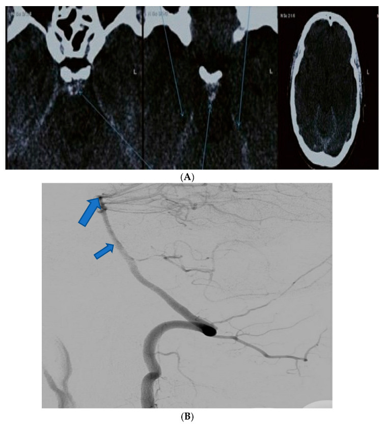
Figure 1.

(A) The case of a 41-year-old male without any significant clinical history who presented in our clinical establishment complaining of diplopia. Axial native CT highlighting blood near the cerebellar tentorium and prepontine cistern (arrows). (B) The same case. Digital subtraction angiography showing no aneurysmal dilatation of the basilary artery (small arrow), cerebellar or proximal posterior cerebral artery (large arrow).
