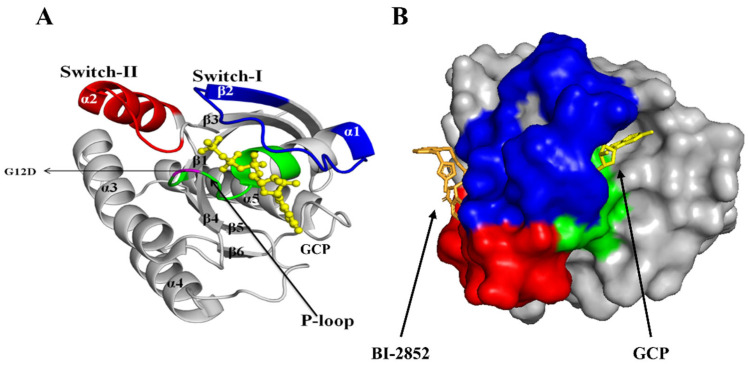
Figure 2.
KRAS G12D mutant protein architecture: (A) A 3-D cartoon view of the GTP-bound KRAS G12D mutant protein (PBD ID: 6GJ8) was visualized, and its α-helices (α1, α2, α3, α4, α5), β-sheets (β1, β2, β3, β4, β5, β6), switches (SI and SII) and P-loop are represented. The missense substitution mutation is present in the 12th position (in the P-loop) where the glycine (G) is substituted by aspartic acid (D). (B) Co-crystallized structure of phosphomethylphosphonic acid guanylate ester (GCP) (yellow) and BI-2852 (bright orange) in KRAS G12D mutant protein (PBD ID: 6GJ8). The P-loop (residues 10–17), switch I (residues 25–40), and switch II (residues 57–76) are shown in green, blue, and red, respectively. The SI/SII binding site of BI-2852 was selected for the molecular-docking studies.

