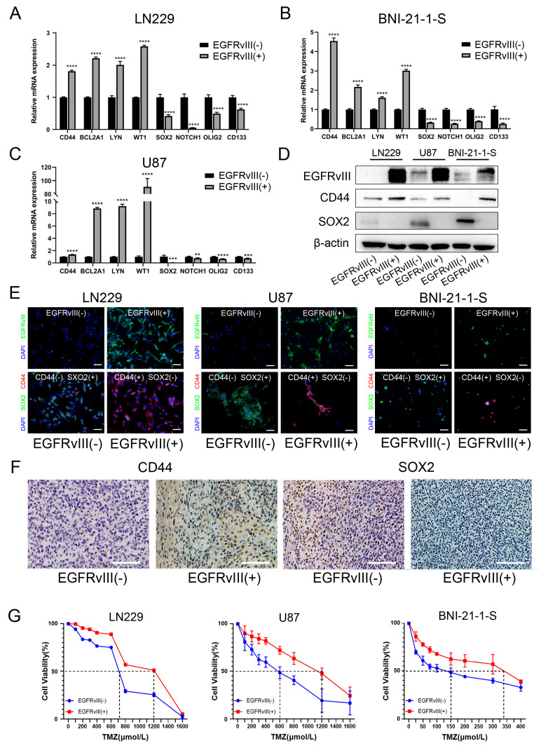
Figure 3.
GBM cells and glioma stem cells with EGFRvIII (+) significantly decreased the expression of PN GBM markers, increased the expression of MES GBM markers, and increased the IC50 value of TMZ. (A–C) The mRNA expression of PMT-related markers were detected by qRT-PCR in U87-EGFRvIII (+) cells, LN229-EGFRvIII (+) cells, and BNI-21-1-S-EGFRvIII (+) cells. (D) The protein expressions of PMT-related markers were detected by Western blot. (E) The expression of PMT-related markers detected by immunofluorescence (green for EGFRvIII and SOX2, red for CD44, blue for DAPI) (scale bars: 50 μm). (F) The expressions of PMT-related markers were detected by immunohistochemistry in tumor tissue from the orthotopic xenograft tumor model of mice (n = 3 per group) (scale bars: 100 μm). (G) The half-maximal inhibitory concentration of U87-EGFRvIII (+) cells, LN229-EGFRvIII (+) cells, and BNI-21-1-S-EGFRvIII (+) cells on TMZ was detected by CCK8. Representative data are determined from three independent experiments (n = 3 per group) vs. EGFRvIII (−). ** p < 0.01, *** p < 0.001, **** p < 0.0001 by two-tailed Student’s t-test.

