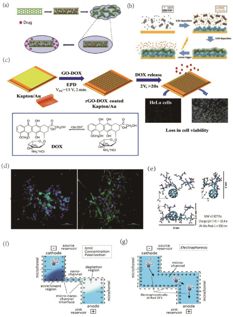
Figure 5.
(a)Schematic of CNT nano reservoirs’ drug loading and release process. Reprinted with permission from [112] Copyright © 2023, Elsevier. (b) Illustration of the synthesis of dual−layer PEDOT/fCNT−PPy/fCNT/DNQX film and controlled release of DNQX from the film. Reprinted with permission from [113] Copyright © 2023, Wiley. (c) Schematic representation of the formation of Au/Kapton flexible electrodes coated with doxorubicin−loaded reduced graphene oxide (rGO-DOX) upon application of a VDC = 15 V for 2 min followed by the electrochemically triggered desorption of the drug and the in vivo test of DOX activity on HeLa cells. Reprinted with permission from [115] Copyright © 2023, The Royal Society of Chemistry. (d) Immunofluorescence staining was performed in NSCs after seven days of differentiation. The markers are green, while the cell nuclei, counterstained with DAPI, are blue. All scale bar lengths are 100 μm. Reprinted with permission from [116] Copyright © 2023, The Royal Society of Chemistry. (e) The structure and properties of DF−1. Schematic of nanochannel Delivery System (nDS) membrane under (f) ionic concentration polarization (ICP) effect in a slit nanochannel and (g) electrophoretic effect in a microchannel. Reprinted with permission from [119] Copyright © 2023, The Royal Society of Chemistry.

