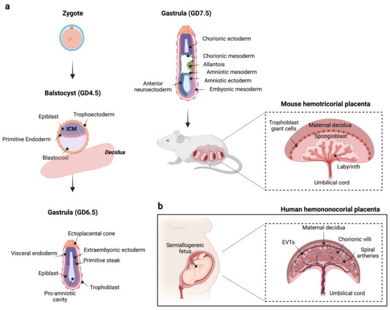
Figure 1.
(a) A schematic representation of mouse placenta development. At gestational day (GD) 3.5–5.0, the mouse blastocyst is composed by ICM, which contains precursor cells for embryonic tissues (epiblast), and visceral and parietal endoderm (primitive endoderm), and by an outer cell layer called trophectoderm. Starting from GD 5.0–6.5, the polar trophectoderm differentiates in the ectoplacental cone, which gives origin to invading trophoblasts and junctional zone, and in the extraembryonic trophectoderm, which, in turn, gives rise to the chorionic ectoderm (GD 6.5–9.5). At the same time, the mesoderm (primitive streak) emerges between visceral endoderm and ectoderm tissues. Subsequently, the chorion fuses with mesodermal-derived allantois promoting placental labyrinth formation. As pregnancy progresses, a definitive placental structure becomes evident around the second week of gestation (GD 10–14.5). (b) A schematic representation of human placenta architecture. Figure created with “https://www.biorender.com/ (accessed on 18 January 2023)”.

