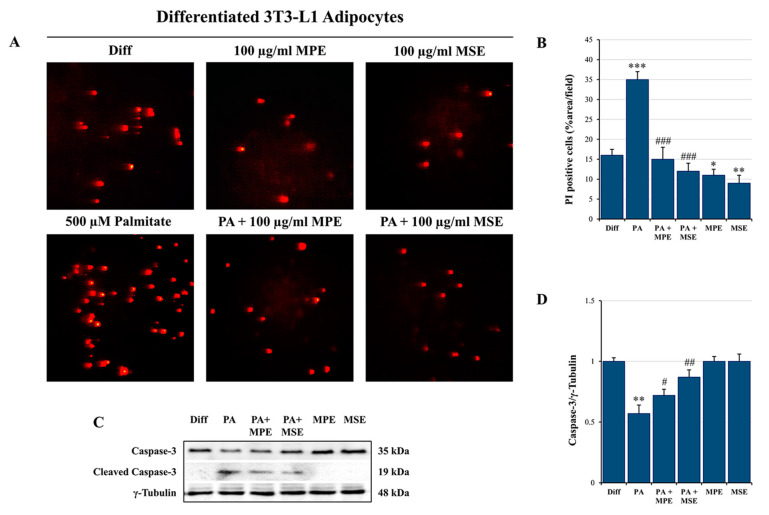
Figure 7.
MPE and MSE reduce the cytotoxic effects of PA in 3T3-L1 adipocytes. Propidium iodide (PI) staining of differentiated 3T3-L1 adipocytes treated with 500 µM PA for 48 h in the presence or absence of 100 µg/mL MPE or MSE. (A) Representative fluorescence microscopy images were taken at 200× magnification by a Leica microscope equipped with a DC300F camera using a PE filter. (B) PI content was ascertained by analyzing the percentage area with Image J. (C) Western blotting analysis of the procaspase-3 levels. An equal loading of protein (30 µg) was verified by immunoblotting for γ-Tubulin (D). The bar graphs represent the means of three independent experiments ± SD. * p < 0.05, ** p < 0.01 and *** p < 0.001 with respect to differentiated 3T3-L1 adipocytes. # p < 0.05, ## p < 0.01, ### p < 0.001 were significant with respect to PA-treated 3T3-L1 adipocytes.

