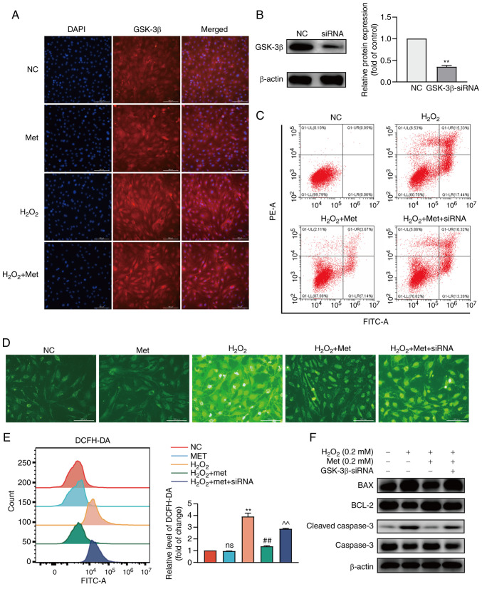
Figure 3.
Metformin improves pre-osteoblast apoptosis induced by oxidative stress through GSK-3β. (A) The localization and expression of GSK-3β was observed in MC3T3-E1 cells and GSK-3β was in a more aggregated state. (B) The silencing efficiency of GSK-3β was detected at the protein level. (C) The apoptotic rate after the silencing of GSK-3β was detected. The apoptotic rate of the H2O2 group was 32.77%, that of the H2O2 + metformin group was 10.81%, and that of the H2O2 + metformin + siRNA group was 23.52%. (D) The level of ROS in the cytoplasm was detected before and after treatment with metformin and siRNA-GSK-3β. (E) The fluorescence intensity of ROS was detected using flow cytometry. (F) The expression of mitochondrial apoptosis-related proteins after the silencing of GSK-3β was detected again. The experiments were performed three times. Data are presented as the mean ± SD. **P<0.01, compared with the control cells; ##P<0.01, compared with the H2O2 group; ^^P<0.01, compared with the H2O2 + Met group. ns, not significant; ROS, reactive oxygen species; Met, metformin; H2O2, hydrogen peroxide; GSK-3β, glycogen synthase kinase 3β.

