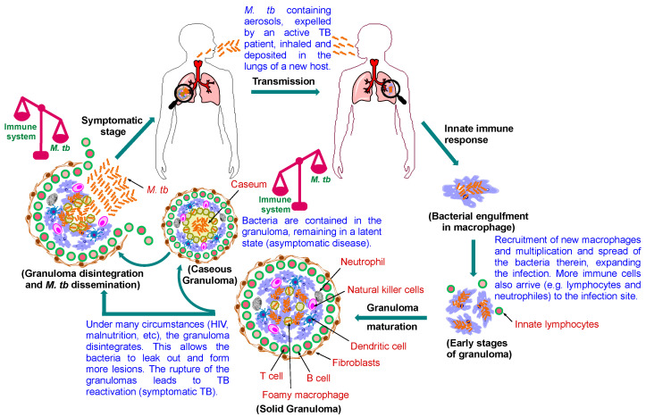
Figure 2.
Pathophysiology of pulmonary TB. Following the M. tb transmission to the new host, the bacilli enter the lung and get ingested by macrophages. Further immune cells are recruited to wall off the infected macrophages, leading to the formation of the granuloma, the hallmark of TB. Healthy individuals remain latently infected, and the infection is kept at bay at this stage, but it is prone to the risk of reactivation. Foamy macrophages release their lipid content when they necrotise, leading to caseation (cheese-like structure). Caseum is a decay manifested at the core of the granuloma that compromise its rigid integrity. As the granuloma develops, the bacilli commence to seep out of the macrophages into the caseum layer. When the reactivation occurs, M. tb proliferates and the bacterial load becomes overwhelmingly high, whereupon the granuloma rupture, disseminating the bacteria to the airways. The bacilli are then expectorated as contagious aerosol droplets, restarting the cycle, infecting other individuals.

