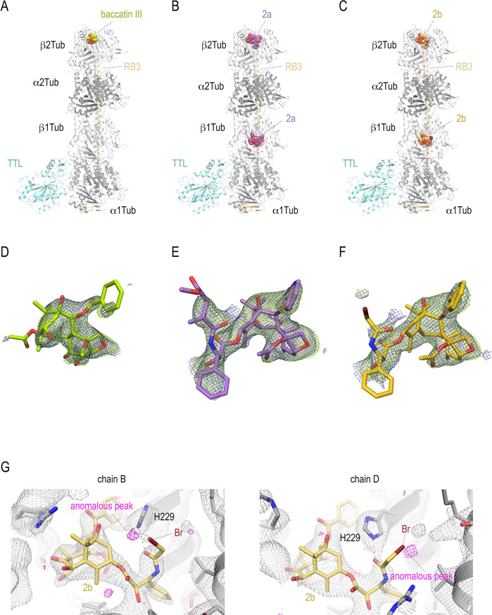
Figure 2. T2R-TTL structures in complex with baccatin III, 2a, and 2b.
Overall view of the T2R-TTL-baccatin III (PDB ID 8BDE) (A), the T2R-TTL-2a (PDB ID 8BDF) (B), and the T2R-TTL-2b (PDB ID 8BDG) crystal structures. The α- and β-tubulin chains are colored in dark and light gray, respectively. The TTL chains (cyan) and the RB3 (yellow-orange) are shown in ribbon representation. The tubulin-bound ligands are displayed as spheres and are colored following the same color scheme as in the main figures. (D–F) Electron-density maps highlighting the bound baccatin III, 2a, and 2b. The SigmaA-weighted 2mFo − DFc (dark blue mesh) and mFo − DFc (light green mesh) omit maps are contoured at +1.0σ and +3.0σ, respectively. The map calculations excluded the atoms of the corresponding ligands. (G) Anomalous density peaks detected in both the binding sites in chains B and D of T2R-TTL for the bromine moiety of compound 2b.

