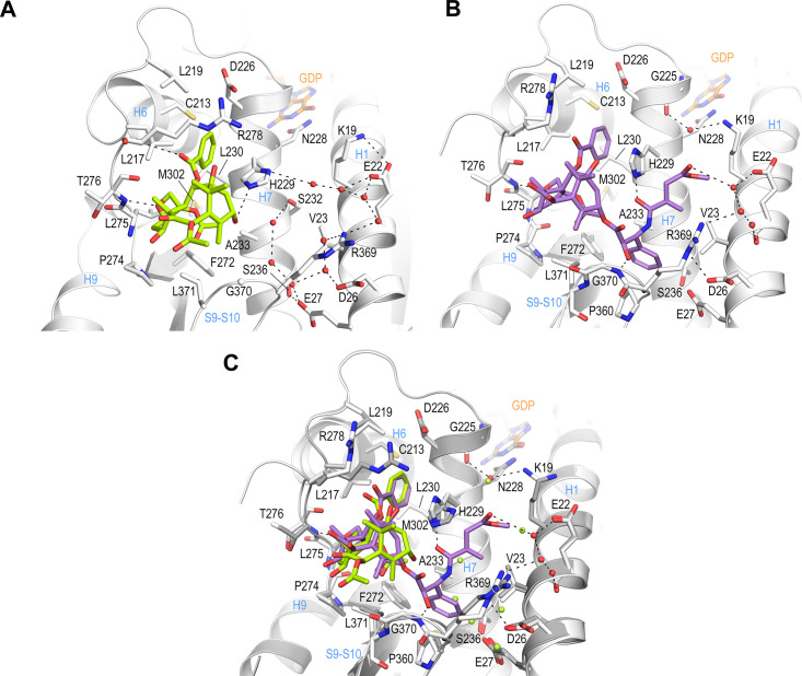
Figure 3. Crystal structure of T2R-TTL-baccatin III (PDB ID 8BDE) and T2R-TTL-2a (PDB ID 8BDF) complexes.
(A) Close-up view of the interaction network observed between baccatin III (lemon) and β-tubulin (light gray). Interacting residues of tubulin are shown in stick representation and are labeled. Oxygen and nitrogen atoms are colored red and blue, respectively; carbon atoms are in lemon (baccatin III) or light gray (tubulin). Hydrogen bonds are depicted as black dashed lines. Secondary structural elements of tubulin are labeled in blue. (B) Close-up view of the interaction of 2a (violet) with β-tubulin in the same view and representation as in (A). (C) The same close-up view as in (A) and (B) with the superimposed baccatin III (lemon) and 2a (violet) complex structures. Water molecules belonging to the baccatin III structure are represented as lemon spheres.

