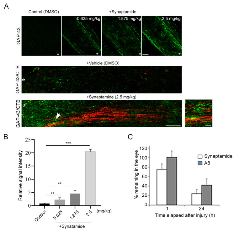
Figure 1.
Intravitreal injection of synaptamide stimulates axon regeneration and extension after injury. (A). Synaptamide dose-dependent axon regeneration indicated by GAP43 staining of optic nerve (top), and synaptamide (2.5 mg/kg)-induced GAP43-positive regenerating axons (green) and axonal extension indicated by CTB labeling (red) at 3 weeks after ONC (bottom) compared to the vehicle-treated control (middle). Longitudinal sections through the optic nerve were collected at 3 weeks after ONC and regenerating axons were visualized by immunostaining for GPA-43, a regenerating axon marker (top). On the third day prior to euthanasia, the animals were injected with CTB-Alexa Fluor 555-conjugated cholera toxin subunit B (CTB, red) as an anterograde tracer to visualize axons in the optic nerve originating from RGCs (bottom). Lesion sites were marked by asterisks (*). Scale bar, 100 μm. An enlarged view around the white arrow is shown as the boxed area, indicating overlapping signals of GAP-43 and CTB. Scale bar, 20 μm. (B). The fluorescent intensities of GAP-43 staining shown in ((A) top) were quantified by setting the whole captured image as the region of interest. The signal intensity was shown relative to the control group. Data are expressed as mean ± s.e.m. (n = 3 per group). ** p < 0.01, *** p < 0.001. (C). The time course of synaptamide and A8 detected in the eye. Synaptamide (2.5 mg/kg) or A8 (0.3 mg/kg) intravitreally injected were detected by tandem mass spectrometry. The data are expressed as mean ± SD (n = 3).

