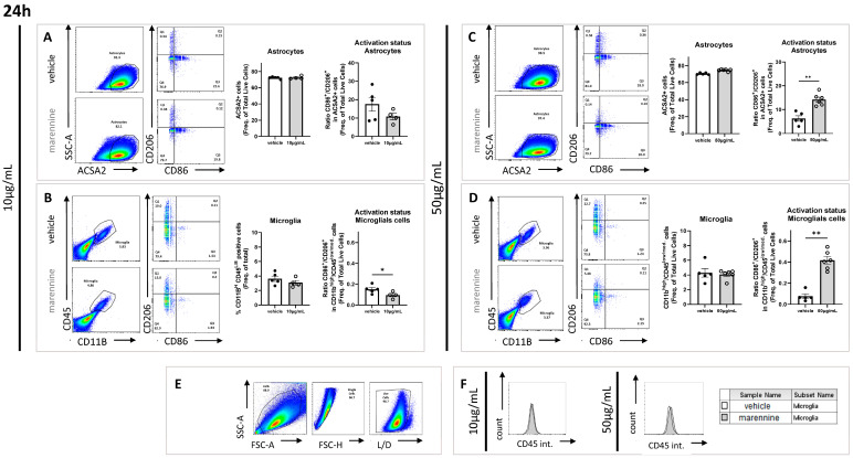
Figure 3.
Effects of 10 and 50 µg/mL of marennine on astrocytes and microglial cells after 24 h of exposure. (A) Astrocytes were gated as ACSA2 positive; activation status was determined through CD86 and CD206 staining after exposure to 10 µg/mL marennine. (B) Microglial cells were gated as CD11bhigh and CD45low/medium; activation status was determined through CD86 and CD206 staining after exposure to 10 µg/mL marennine. (C) Astrocytes were gated as ACSA2 positive; activation status was determined through CD86 and CD206 staining after marennine exposure at 50 µg/mL. (D) Microglial cells were gated as CD11bhigh and CD45low/medium; activation status was determined through CD86 and CD206 staining after marennine at 50 µg/mL. (E) Cell populations and activation status were determined by flow cytometry. A gate was first created on non-debris populations, excluding very low SSC/FSC events; cells are then gated on single cells and selected by Live/Dead staining. (F) Quantification of CD45 intensity after a marennine of 10 µg/mL (left) and 50 µg/mL (right). For 24 h, at 10 µg/mL vehicle n = 5, marennine n = 4. At 50 µg/mL vehicle n = 5, marennine n = 6. Values represent mean ± SEM. Significant differences from control are indicated as follows: * p < 0.05; ** p < 0.01 using Mann-Whitney test.

