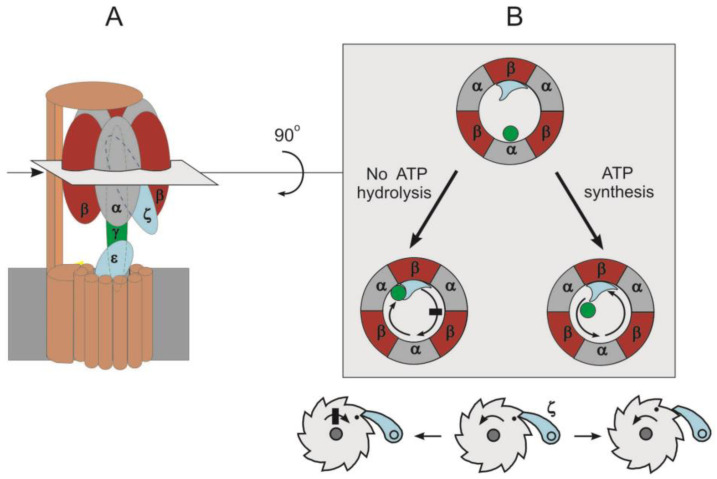
Figure 2.
The inhibition of ATP hydrolysis in F1·Fo by inhibitor protein according to pawl-ratchet mechanism. (A): Schematic representation of F1·Fo (P. denitrificans). The subunit colors are the same as in Figure 1, but the peripheral stalk and c-ring are shown in brown and ζ subunit is colored in blue. The deep insertion of inhibitory protein (ζ subunit) into αβ interface is depicted. Cross-section through the F1 domain is shown. (B): View of F1 from the top; for clarity, the δ and bb1 subunits are not shown. Β and ζ subunits form a small indentation acting as pawl teeth. The pawl-ratchet mechanism enables the γ subunit’s free rotation in the ATP synthesis direction but stops rotation in ATP hydrolysis direction.

