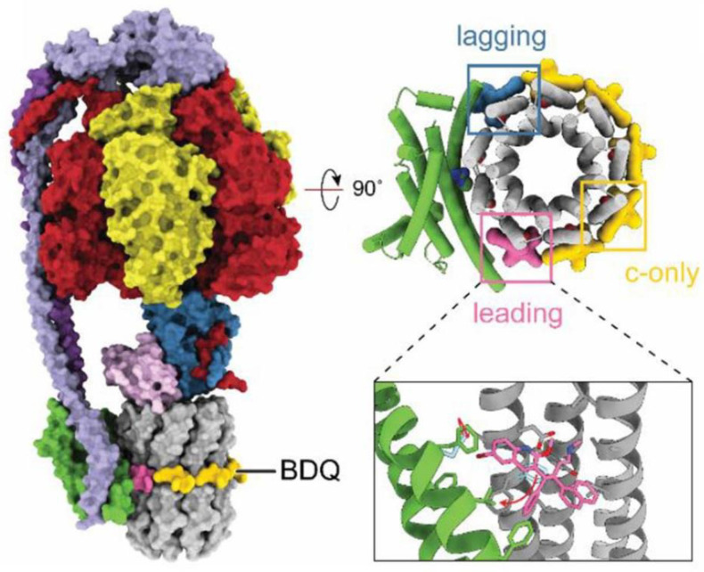
Figure 5.
Structure of M. smegmatis F1·Fo ATP synthase bound to bedaquiline (PDB ID: 7JGC). Bedaquiline (BDQ) binds at five c-only sites (yellow), a leading site (pink), and a lagging site (blue) in the Fo region of the enzyme. Red arrows show the movement of residues upon bedaquiline binding. Adapted from Courbon and Rubinstein [6] under the terms of the Creative Commons Attribution 4.0 International Public License.

