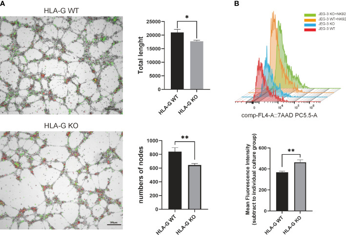
Figure 4.
HLA-G functional assay on the JEG-3 cells co-cultured model. (A) Images and representative statistical bar graphs of an angiogenesis assay in Jeg-3 cells (green) transfected with HLA-G knockout or empty vectors under a 24 h co-culture with HUVEC cells (red). (B) Colored histogram (left) and mean fluorescence intensity (right) of 7AAD staining (PC5.5-A) results with cell viability between JEG-3 KO and WT cells in co-culture with NK92 cells. *P<0.05 or **P<0.01. n = 3 in triplicate.

