FIGURE 1.
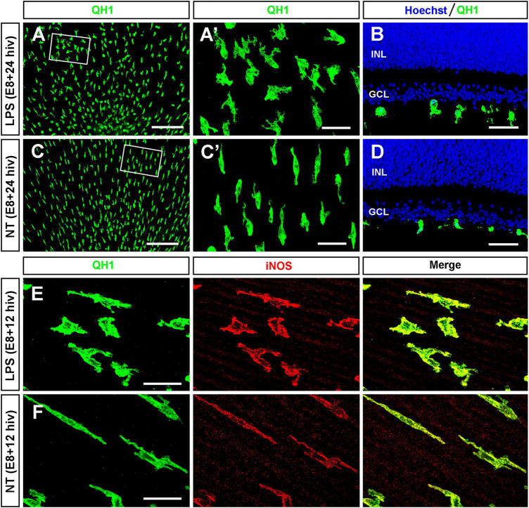
LPS-treatment of E8 retina explants induces changes in morphology and iNOS expression in immature microglia. (A–D) Representative images of QH1-immunolabeled microglia (green) in whole-mounts (A,C) and cross cryosections (B,D) of LPS-treated [LPS; (A,B)] and non-treated [NT; (C,D)] retina explants from E8 quail embryos cultured for 24 h (E8 + 24 hiv). Hoechst nuclear staining (blue) was used to visualize the retinal layers in cryosections (INL: inner nuclear layer; GCL: ganglion cell layer). The boxed areas in panels (A,C) are shown at a higher magnification in panels (A’,C’), respectively. Note morphological changes of microglial cells in LPS-treated explants (A,A’,B) compared with those in the non-treated explants (C,C’,D). (E,F) Representative confocal images of QH1 (green) and anti-iNOS (red) double immunostained microglial cells in LPS-treated [LPS; (E)] and non-treated [NT; (F)] E8 + 12 hiv retina explants. Green, red, and merged channels are shown in the left (QH1), medium (iNOS), and right (Merge) panels, respectively. In addition to that, morphological changes similar to those found in E8 + 24 hiv retina explants, the iNOS labeling of microglia is higher in LPS-treated versus non-treated explants. Scale bars: 200 μm in panels (A,C); 40 μm in panels (A’,B,C’,D); 25 μm in panels (E,F).

