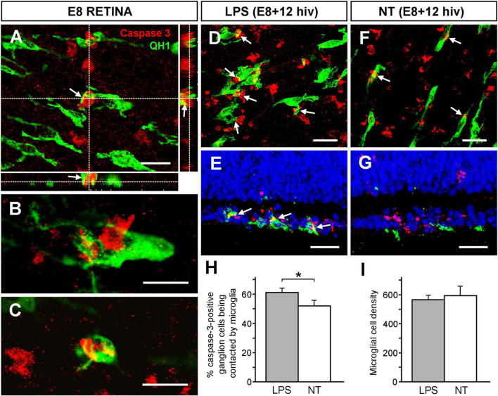
FIGURE 4.
Increase of phagocytic contacts between microglial cells and caspase-3-positive ganglion cells. The frequency of phagocytic contacts between microglial cells and caspase-3-positive ganglion cells increases in response to LPS treatment in retina explants from E8 quail embryos cultured for 12 h in vitro (E8 + 12 hiv). (A–C) Confocal images of microglial cells forming close engulfing contacts with caspase-3-positive ganglion cells in whole-mounted non-cultured E8 quail embryo retina explants, (E8 RETINA) double immunolabeled with QH1 (green) and anti-caspase-3 (red). These images are maximum-intensity projections from Z stacks of confocal optical sections throughout the thickness of the ganglion cell layer (GCL). Panel (A) shows a process from a microglial cell that contacts and embraces a caspase-3-positive ganglion cell. The XZ and YZ orthogonal projections corroborate the close contact (arrows) between the microglial cell process and the ganglion cell. Scale bar: 15 μm. (B) Phagocytic contacts between a microglial cell soma and a caspase-3-positive ganglion cell are observed. (C) A caspase-3-positive ganglion cell completely engulfed by a microglial cell. Scale bars for panels (B,C): 15 μm. (D–G) Representative confocal images of whole-mounts (D,F) and cross-sections (E,G) from LPS-treated [LPS; (D,E)] and non-treated [NT; (F,G)] E8 + 12 hiv retina explants double immunolabeled with QH1 (green) and anti-caspase-3 (red) showing phagocytic contacts (arrows) between microglial cells and caspase-3-positive ganglion cells. Confocal images of whole-mounted explants are optical sections obtained in the ganglion cell layer. Cell nuclei in cross-sections were stained with Hoechst 33342 (blue) to visualize the retinal layers. Phagocytic contacts between microglial cells and caspase-3-positive ganglion cells are more abundant in LPS-treated explants compared to non-treated ones. Scale bars: 25 μm. (H) Quantification of caspase-3-positive ganglion cells that are contacted by microglia in LPS-treated (grey bar) and non-treated (white bar) E8 + 12 hiv retina explants. Bars represent means (±SEM) of percentages obtained from counts in three different zones (dorsal, nasal and temporal) of whole-mounted explants (n = 6 for each condition). The percentage of caspase-3-positive ganglion cells being contacted by microglia is significantly higher in LPS-treated than in non-treated explants (*P < 0.05, Student’s t-test). (I) Microglial cell densities in LPS-treated (LPS, grey bar) and non-treated (NT, white bar) E8 + 12 hiv retina explants. Data are presented as means (±SEM) of microglial cells per mm2 obtained from counts in three square fields of 500 μm × 500 μm per whole-mounted retina explant (n = 10 for each condition). No significant differences are detected between LPS-treated and non-treated explants.

