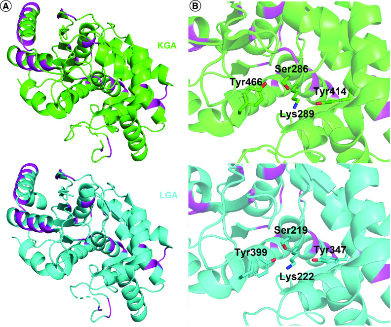
Figure 3. . GLS and GLS2 share highly conserved active sites.
(A) Alignment of the catalytic domains of KGA (green, PDB: 3czd) and LGA (cyan, PDB: 4bqm). The few differences are highlighted in magenta. (B) A closer look at the substrate-binding pocket, with the catalytically active residues shown as sticks.

