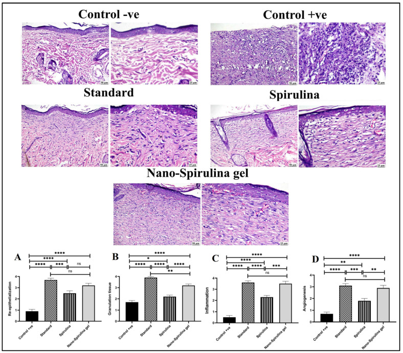
Figure 12.
Photomicrographs of skin (H&E) stained showing normal skin in the -ve control group, the absence of epithelial cover and intense inflammation in the +ve control group, good re-epithelialization and collagen-rich granulation tissue in the standard group, complete re-epithelialization, good granulation tissue formation and mild inflammation in the S. platensis gel group, perfect wound closure, re-epithelialization, and minimal inflammation in the nano-S. platensis gel (SPNP-gel) group. The charts describe the histopathological scores of wound healing criteria in different experimental groups. (A) Re-epithelization score, (B) Granulation tissue formation, (C) Inflammation score and (D) Angiogenesis. Control -ve group showed normal skin (no healing criteria could be evaluated). Data were presented as means ± SD, and a significant difference was considered at p ˂ 0.05. Note: GpI/control negative (-ve): non-wounded rats; GpII/control positive (+ve): untreated wounded rats; GpIII/standard group: wounded rats treated with MEBO®; GpIV/S. platensis gel: wounded rats treated with S. platensis gel; and GpV/nano-spirulina (SPNP)-gel: wounded rats treated with SPNP-gel. Number of asterisks “*” above the columns indicates strength of significance, (ns) means non-significant.

