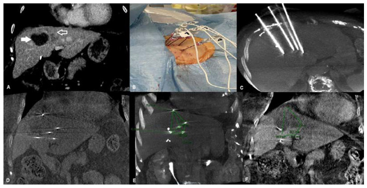
Figure 1.
An 81-year-old female affected by recurrence of liver metastasis from pancreatic cancer after percutaneous ablation and SBRT, treated with percutaneous ECT. (A) Pre-treatment coronal CT reconstruction showing dishomogeneous enhancing tissue consistent with metastatic liver recurrence (contoured arrow) at the periphery of the hypodense post-ablation and post-SBRT area (white arrow). (B) Percutaneous placement of the five electrodes. (C) Unenhanced cone-beam CT MPR and MIP reconstruction showing the five electrodes positioned in the target lesion. (D) Unenhanced cone-beam CT coronal reconstruction showing the tip of the five electrodes in place. (E) Unenhanced cone-beam CT coronal reconstruction showing the tip of the five electrodes in place and the post-processing measurements underlining the geometrical relationship between each electrode. (F) Contrast-enhanced post-ECT cone-beam CT coronal reconstruction showing the ablation area. Legend: ECT: Electrochemotherapy; SBRT: Stereotactic body radiation therapy; CT: Computed tomography; MPR: Multiplanar reconstruction; MIP: Maximum intensity projection.

