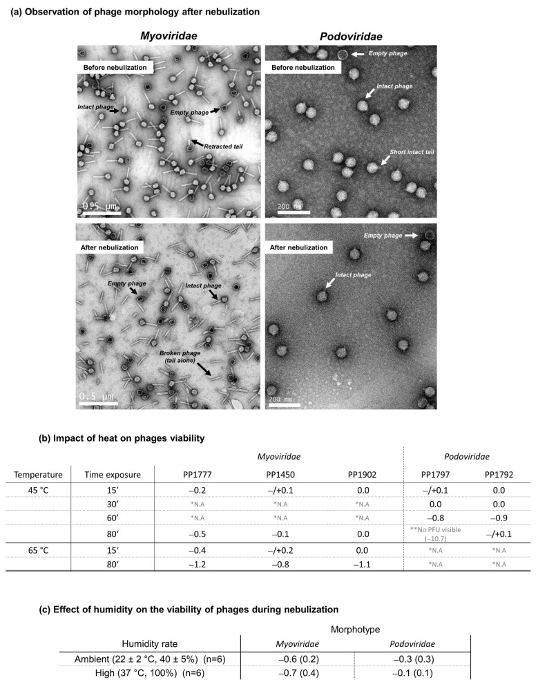
Figure 6.
Impact of nebulization on phage morphology and impacts of heat and humidity during nebulization on phage viability. (a) Observation of phage morphology after nebulization: Representative Transmission Electron Micrographs of Myoviridae and Podoviridae before and after nebulization with the prototype static-mesh nebulizer. The white scale lines correspond to 0.5 µm for Myoviridae and 200 nm for Podoviridae. The white or black arrows show and name the morphological structures observed. Intact phage is considered as a phage with its head, its genome and its tail. (b) Impact of heat on phage viability: For each time exposure, the phage viability was expressed as a loss of log PFU, based on the difference between the log PFU measured at 45 °C or at 65 °C with the log PFU measured at 4 °C (mean log PFU45 °C or 65 °C—mean log PFU4 °C). Each value of log PFU was a mean of three replicates. * N.A., for Not Available, means that the time exposure was not tested; ** no PFU visible means that any lysis spot was visible. (c) Impact of humidity on the viability of phages during nebulization: loss of log pfu (mean ± SD) after nebulization under ambient humidity (22 °C, 40%) or high humidity rate (37 °C, 100%).

