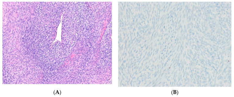
Figure 6.
Malignant peripheral nerve sheath tumor. (A) The tumor shows spindle cells in a fascicular pattern. Perivascular accentuation of cellularity is present (H&E stain, ×100). (B) The tumor cells show loss of H3K27me3 expression, indicating the presence of underlying inactivating mutations of the polycomb repressive complex 2 components (SUZ12 or EED) (immunohistochemical stain for H3K27me3, ×200).

