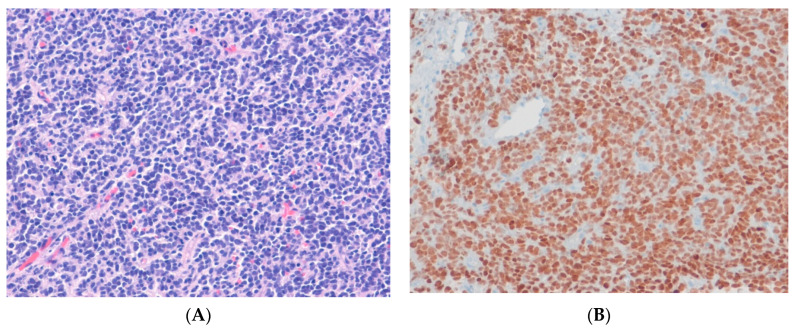
Figure 9.
BCOR-rearranged sarcoma. (A) The tumor shows solid sheets of monomorphic round cells with round nuclei and scant eosinophilic cytoplasm, with delicate capillaries (H&E stain, ×100). (B) The tumor cells show diffuse nuclear staining for BCOR, indicating the presence of an underlying BCOR::CCNB3 fusion gene (immunohistochemical stain for BCOR, ×200).

