Figure 5.
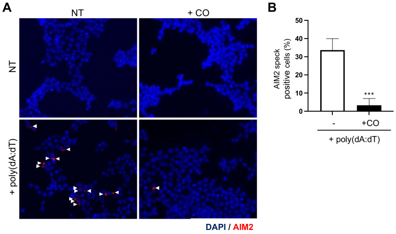
CO inhibits AIM2 speck formation resulting from poly(dA:dT) stimulation. (A,B) HEK293T cells were transfected with a FLAG-tagged AIM2 expression vector 24 h prior to the experiment. Cells were pre-treated with CO (10 μg/mL) for 30 min, then stimulated with poly(dA:dT) (1 μg/mL) for 4 h. (A) AIM2 speck images at 400× magnification upon stimulation with poly(dA:dT) in cells expressing FLAG-tagged AIM2. AIM2 and nuclei are depicted in red and blue (DAPI), respectively. White arrowheads point to AIM2 speck. (B) Statistical analysis of the AIM2 specks showing the percentage of positive cells. At least three fields and more than 100 cells were counted for each condition. Data represent the means ± SDs of three independent experiments; *** p < 0.001.

