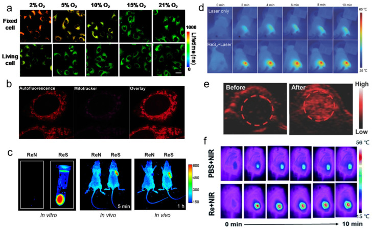
Figure 9.
(a) PLIM images of Re-14a-treated fixed and living A549 cells under different oxygen partial pressures at 37 °C (reproduced with permission from Ref. [108], Copyright 2017, American Chemical Society); (b) magnified images showing an overlay of Cp-Dox autofluorescence and MitoTracker fluorescence in HeLa cells, suggesting an accumulation of the Dox conjugate in the mitochondrial membrane (reproduced with permission from Ref. [114], Copyright 2016, Wiley-VCH); (c) fluorescence emission of Re-27a and Re-27b in vitro and in vivo (reproduced with permission from Ref. [107], Copyright 2019, American Chemical Society); (d) thermal images of PVP capped ReS2 in in vivo PTT (reproduced with permission from Ref. [132], Copyright 2018, Wiley-VCH); (e) PA images of tumors in mice before and 24 h after i.v. injection of ReS2-PEG (reproduced with permission from Ref. [133], Copyright 2017, Wiley-VCH); (f) thermal imaging of tumor-bearing mice after injection of PBS or Re NCs (reproduced with permission from Ref. [131], Copyright 2010, Royal Society of Chemistry). PLIM, phosphorescence lifetime imaging; PVP, polyvinyl pyrrolidone; PTT, photothermal therapy; PA, photoacoustic; PEG, polyethylene glycol; PBS, phosphate buffer saline; NCs, nanoclusters.

